WHAT IS A STROKE?
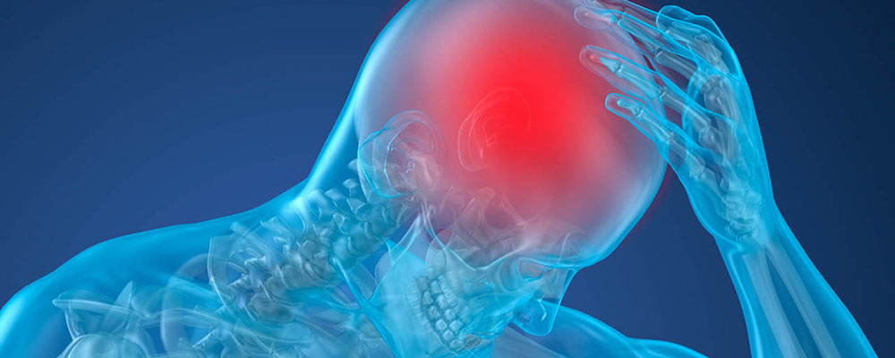
A stroke occurs when a blood vessel that carries oxygen and nutrients to the brain is either blocked by a clot or bursts (or ruptures). When that happens, part of the brain cannot get the blood (and oxygen) it needs. This results in brain tissue damage and brain cell death. Thus, disrupting and/or causing loss of certain brain functions

CLASSIFICATION OF STROKE
There are 2 main types of stroke:
- Ischemic stroke is the most common type of stroke, accounting for about 80% of all stroke cases. This can be an obstruction caused by a blood clot or piece of debris that forms in another area and travels along the bloodstream until it causes a blockage of an artery in the brain. An ischemic stroke can also be the result of the accumulation of fatty deposits and cholesterol in the arterial wall, which causes narrowing of the blood vessels and loss of flexibility, resulting in an inefficient transport of oxygen-rich blood to the brain.
- Hemorrhagic stroke accounts for about 20% of stroke cases. The most common causes of hemorrhagic stroke are high blood pressure and brain aneurysms, as the weak or bulging area in the arterial wall balloons and eventually ruptures. It can also be caused by artery weakening or loss of flexibility due to accumulation of fat in the arteries, making them susceptible to breakage and rupture. This is very dangerous as it causes a sudden drop in blood supply to the brain, as well as bleeding within the brain, which can result in death in a very short period of time.
Transient Ischemic Attack (TIA) is a temporary blockage of blood flow to the brain. The clot usually dissolves on its own or gets dislodged, and the symptoms usually last less than five minutes. While a TIA does not cause permanent damage, it is a “warning stroke” signalling a possible full-blown stroke ahead. When you first notice symptoms, get help immediately, even if symptoms go away.
Brain Stem Stroke The brain stem controls all basic activities of the central nervous system: consciousness, blood pressure and breathing. All motor control for the body flows through it. When stroke occurs in the brain stem, it can affect both sides of the body and may leave a person in a “locked-in” state. When a locked-in state occurs, the patient is generally unable to speak or move below the neck. More severe brain stem strokes can cause locked-in syndrome, a condition in which survivors can move only their eyes
NON-MODIFIABLE AND MODIFIABLE RISKS
There are a variety of factors which can increase the risk of a stroke. These are divided into non-modifiable risk factors and modifiable risk factors.
Non-Modifiable Risks
- Age — as a person gets older, their blood vessels weaken and deteriorate, whereby the inner layer of the artery walls thicken and harden due to plaque buildup, causing a gradual narrowing of the arteries.
- Gender — men have a higher risk of stroke than women.
- Hypercoagulable States — this condition causes blood platelets to stick together and clot more easily than normal.
Modifiable Risks
- High blood pressure is the most important risk factor for stroke. People who have high blood pressure are at much greater risk of stroke.
- Diabetes causes hardening of the arteries throughout the body. If this occurs in the brain, it increases the chance of stroke by 2-3 times.
- High cholesterol is just as a risk factor for stroke as it is for coronary artery disease. The accumulation of fatty deposits in the arterial wall results in narrowing of lumen, making it difficult for an efficient amount of blood to flow through.
- Heart disease, such as atrial fibrillation and arrhythmia, causes blood clots. If a blood clot forms in an artery that supplies blood to the brain, it could result in a blood deficiency in the brain.
- Smoking is a risk factor, as nicotine and carbon monoxide lower the oxygen levels in the blood, damage the arterial walls, and causes hardening of the arteries.
- Oral contraceptives are a risk factor, as the chance of a stroke is higher in women using birth control pills with high doses of estrogen.
- Syphilis can cause inflammation of cerebral blood vessels and hardening of the arteries.
- Physical inactivity and lack of exercise raises the risk of stroke.
SIGNS & SYMPTOMS OF STROKE
F.A.S.T. Warning Signs
Use the letters in F.A.S.T. to spot a Stroke
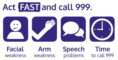
- F = Face Drooping – Does one side of the face droop or is it numb? Ask the person to smile. Is the person's smile uneven?
- A = Arm Weakness – Is one arm weak or numb? Ask the person to raise both arms. Does one arm drift downward?
- S = Speech Difficulty – Is speech slurred?
- T = Time to seek for treatment – Stroke is an emergency. Every minute counts. Call for ambulance or proceed to the nearest hospital. Note the time when any of the symptoms first appear

Other Stroke Symptoms
Watch for Sudden:
- NUMBNESS or weakness of face, arm, or leg, especially on one side of the body
- CONFUSION, trouble speaking or understanding speech
- TROUBLE SEEING in one or both eyes
- TROUBLE WALKING, dizziness, loss of balance or coordination
- SEVERE HEADACHE with no known cause
If any of these abnormal symptoms occur, it is vital that patients seek immediate medical attention. As a stroke can be severe and potentially life-threatening, the outcome depends on how soon treatment starts.
EFFECTS OF STROKE
The effects of a stroke depend on several factors, including the location of the obstruction and how much brain tissue is affected. However, because one side of the brain controls the opposite side of the body, a stroke affecting one side will result in neurological complications on the side of the body it affects
|
LEFT BRAIN |
RIGHT BRAIN |
|---|---|
|
If the stroke occurs in the left side of the brain, the right side of the body will be affected, producing some or all of the following:
|
If the stroke occurs in the right side of the brain, the left side of the body will be affected, producing some or all of the following:
|
DIAGNOSTIC TEST
Current diagnostic methods are highly effective and are able to identify the location of the damage or abnormalities in the brain or blood vessels, as well as any conditions and causes that could be risk factors for stroke.
Methods include:
- A physical exam.Your doctor will do a number of tests you're familiar with, such as listening to the heart and checking the blood pressure. You'll also have a neurological exam to see how a potential stroke is affecting your nervous system.
- Blood tests.You may have several blood tests, including tests to check how fast the blood clots, whether the blood sugar is too high or low and also the cholesterol level.
- Computerized tomography (CT) scan.A CT scan uses a series of X-rays to create a detailed image of your brain. A CT scan can show bleeding in the brain, an ischemic stroke, a tumour or other conditions. Doctors may inject a dye into your bloodstream to view the blood vessels in the neck and brain in greater detail (computerized tomography angiography).
- Magnetic Resonance Imaging (MRI).An MRI uses powerful radio waves and a magnetic field to create a detailed view of the brain. An MRI can detect brain tissue damaged by an ischemic stroke and brain haemorrhages. Your doctor may inject a dye into a blood vessel to view the arteries and veins and highlight blood flow (magnetic resonance angiography or magnetic resonance venography).
- Carotid ultrasound.In this test, sound waves create detailed images of the inside of the carotid arteries in the neck. This test shows buildup of fatty deposits (plaques) and blood flow in the carotid arteries.
- Cerebral angiogram.In this uncommonly used test, the doctor inserts a thin, flexible tube (catheter) through a small incision, usually in the groin, and guides it through the major arteries and into the carotid or vertebral artery. Then your doctor injects a dye into the blood vessels to make them visible under X-ray imaging. This procedure gives a detailed view of arteries in the brain and neck.
- An echocardiogram uses sound waves to create detailed images of the heart. An echocardiogram can find a source of clots in the heart that may have travelled from the heart to the brain and caused a stroke.
TREATMENT
Emergency treatment for stroke depends on whether you are having an ischemic stroke or a stroke that involves bleeding into the brain (haemorrhagic).
Ischemic stroke
To treat an ischemic stroke, doctors must quickly restore blood flow to the brain. This may be done with:
- Emergency IV medication. Therapy with drugs that can break up a clot has to be given within 4.5 hours from when symptoms first started if given intravenously. The sooner these drugs are given, the better. Quick treatment not only improves the chances of survival but also may reduce complications.
- An IV injection of recombinant tissue plasminogen activator (TPA) — also called alteplase (Activase) or tenecteplase (TNKase) — is the gold standard treatment for ischemic stroke. An injection of TPA is usually given through a vein in the arm within the first three hours. Sometimes, TPA can be given up to 4.5 hours after stroke symptoms started.
- This drug restores blood flow by dissolving the blood clot causing the stroke. By quickly removing the cause of the stroke, it may help people recover more fully from a stroke. Your doctor will consider certain risks, such as potential bleeding in the brain, to determine whether TPA is appropriate for you.
Emergency endovascular procedures. Doctors sometimes treat ischemic strokes directly inside the blocked blood vessel. Endovascular therapy has been shown to significantly improve outcomes and reduce long-term disability after ischemic stroke. These procedures must be performed as soon as possible:
- Medications delivered directly to the brain. Doctors insert a long, thin tube (catheter) through an artery in the groin and thread it to the brain to deliver TPA directly where the stroke is happening. The time window for this treatment is somewhat longer than for injected TPA but is still limited.
- Removing the clot with a stent retriever. Doctors can use a device attached to a catheter to directly remove the clot from the blocked blood vessel in the brain. This procedure is particularly beneficial for people with large clots that cannot be completely dissolved with TPA. This procedure is often performed in combination with injected TPA.
The time window when these procedures can be considered has been expanding due to newer imaging technology. Doctors may order perfusion imaging tests (done with CT or MRI) to help determine how likely it is that someone can benefit from endovascular therapy.
Other Procedure
To decrease your risk of having another stroke or transient ischemic attack, your doctor may recommend a procedure to open an artery that is narrowed by plaque. Options vary depending on the situation, but include:
- Carotid endarterectomy. Carotid arteries are the blood vessels that run along each side of the neck, supplying the brain (carotid arteries) with blood. This surgery removes the plaque blocking a carotid artery and may reduce the risk of ischemic stroke. A carotid endarterectomy also involves risks, especially for people with heart disease or other medical conditions.
- Angioplasty and stents. In an angioplasty, a surgeon threads a catheter to the carotid arteries through an artery in the groin. A balloon is then inflated to expand the narrowed artery. Then a stent can be inserted to support the opened artery.
Haemorrhagic stroke
Emergency treatment of haemorrhagic stroke focuses on controlling the bleeding and reducing pressure in the brain caused by the excess fluid. Treatment options include:
- Emergency measures.If you take blood-thinning medications to prevent blood clots, you may be given drugs or transfusions of blood products to counteract the blood thinners' effects. You may also be given drugs to lower the pressure in the brain (intracranial pressure), lower blood pressure, prevent spasms of the blood vessels and prevent seizures
- If the area of bleeding is large, your doctor may perform surgery to remove the blood and relieve pressure on the brain. Surgery may also be used to repair blood vessel problems associated with haemorrhagic strokes. Your doctor may recommend one of these procedures after a stroke or if an aneurysm, arteriovenous malformation (AVM) or other type of blood vessel problem caused the haemorrhagic stroke.
- Surgical clipping.A surgeon places a tiny clamp at the base of the aneurysm to stop blood flow to it. This clamp can keep the aneurysm from bursting, or it can keep an aneurysm that has recently haemorrhaged from bleeding again.
- Coiling (endovascular embolization).Using a catheter inserted into an artery in the groin and guided to the brain, the surgeon will place tiny detachable coils into the aneurysm to fill it. This blocks blood flow into the aneurysm and causes blood to clot.
- Surgical AVM removal.Surgeons may remove a smaller AVM if it's located in an accessible area of the brain. This eliminates the risk of rupture and lowers the risk of haemorrhagic stroke. However, it is not always possible to remove an AVM if it's located deep within the brain, it's large, or its removal would cause too much of an impact on brain function.
STROKE RECOVERY AND REHABILITATION
After emergency treatment, the patient will be closely monitored for at least a day. After that, stroke care focuses on helping you recover as much function as possible and return to independent living. The impact of the stroke depends on the area of the brain involved and the amount of tissue damaged.
If the stroke affected the right side of the brain, your movement and sensation on the left side of the body may be affected. If the stroke damaged the brain tissue on the left side of the brain, your movement and sensation on the right side of the body may be affected. Brain damage to the left side of the brain may cause speech and language disorders.
Most stroke survivors go to a rehabilitation program. Your doctor will recommend the most rigorous therapy program you can handle based on your age, overall health and degree of disability from the stroke. Your doctor will take into consideration your lifestyle, interests and priorities, and the availability of family members or other caregivers.
Rehabilitation may begin before you leave the hospital. After discharge, you might continue your program in a rehabilitation unit of the same hospital, another rehabilitation unit or skilled nursing facility, as an outpatient, or at home.
Every person's stroke recovery is different. Depending on your condition, your treatment team may include:
- Doctor trained in brain conditions (neurologist)
- Rehabilitation doctor (physiatrist)
- Rehabilitation nurse
- Dietitian
- Physical therapist
- Occupational therapist
- Recreational therapist
- Speech pathologist
- Social worker or case manager
- Psychologist or psychiatrist
PREVENTION OF STROKE RECURRENCE
Prevention is always the best treatment, and prevention of stroke includes controlling risk factors.
Tips for stroke prevention:
- Have annual health check-ups in order to check for risk factors. If any are found, patients should quickly begin treatment and have regular doctor visits.
- In cases where risk factors are found that could cause artery stenosis, blockage, or rupture, patients can take anti-coagulant and/or anti-platelet medication – following their doctor’s treatment plan. Medications should not be discontinued on one’s own; patients should seek immediate medical attention if any unusual symptoms develop.
- Control high blood pressure, cholesterol, and blood sugar levels – remaining within a normal range.
- Have a well-balanced diet; avoid salty, sweet, and greasy foods.
- Exercise regularly, at least 30 minutes a day, 3 times per week, and maintaining a healthy weight.
- Quit smoking and avoid alcohol.
- If there are any symptoms indicating inadequate blood supply to the brain, seek immediate medical attention.
REFERENCES:
American Stroke Association accessed at https://www.stroke.org/en/about-stroke/types-of-stroke
Mayo Clinic accessed at https://www.mayoclinic.org/diseases-conditions/stroke/symptoms-causes/syc-20350113
